

On this page
Alexander disease
This condition primarily affects the central nervous system, leading to developmental delays, motor difficulties, and seizures. Alexander disease can present at different ages, with infantile forms typically being more severe than juvenile or adult-onset forms.
Overview
Alexander disease is a rare inherited leukodystrophy, a group of conditions affecting the white matter of the brain. White matter consists of nerve fibres that transmit signals throughout the nervous system, and damage to these structures disrupts normal brain function. The condition is caused by pathogenic variants in both copies of GFAP in affected individuals. GFAP provides instructions for making a protein essential to the structure and function of astrocytes, specialised cells that support nerve cells in the brain.
The condition can present at different life stages, with infantile-onset Alexander disease being the most common and typically the most severe form. Later-onset forms, appearing in childhood, adolescence, or adulthood, tend to progress more slowly and may have different clinical features. Regardless of age of onset, the condition is progressive, meaning symptoms worsen over time. Alexander disease affects both males and females and occurs across all ethnic backgrounds, though exact prevalence figures are not well established due to its rarity.
Symptoms & clinical features
Clinical features vary considerably depending on the age at which symptoms first appear. Infantile-onset Alexander disease, which develops before age two years, typically presents with developmental delays, particularly in achieving motor milestones such as sitting or walking. Affected infants often have an unusually large head (macrocephaly), which may be one of the earliest signs. Seizures are common and may be difficult to control with medication. As the condition progresses, children may develop increasing stiffness in the limbs (spasticity) and difficulties with swallowing and feeding.
Juvenile and adult forms of Alexander disease often present differently, with less prominent developmental delay and macrocephaly. Instead, affected individuals may experience difficulties with speech and swallowing, abnormal eye movements, balance problems, and progressive weakness or stiffness in the limbs. Some people develop symptoms related to disruption of the brainstem and spinal cord, including difficulty controlling bladder and bowel function. The rate of progression varies, with some individuals experiencing relatively stable periods followed by periods of decline.
Video: Genetics 101
Affected organs
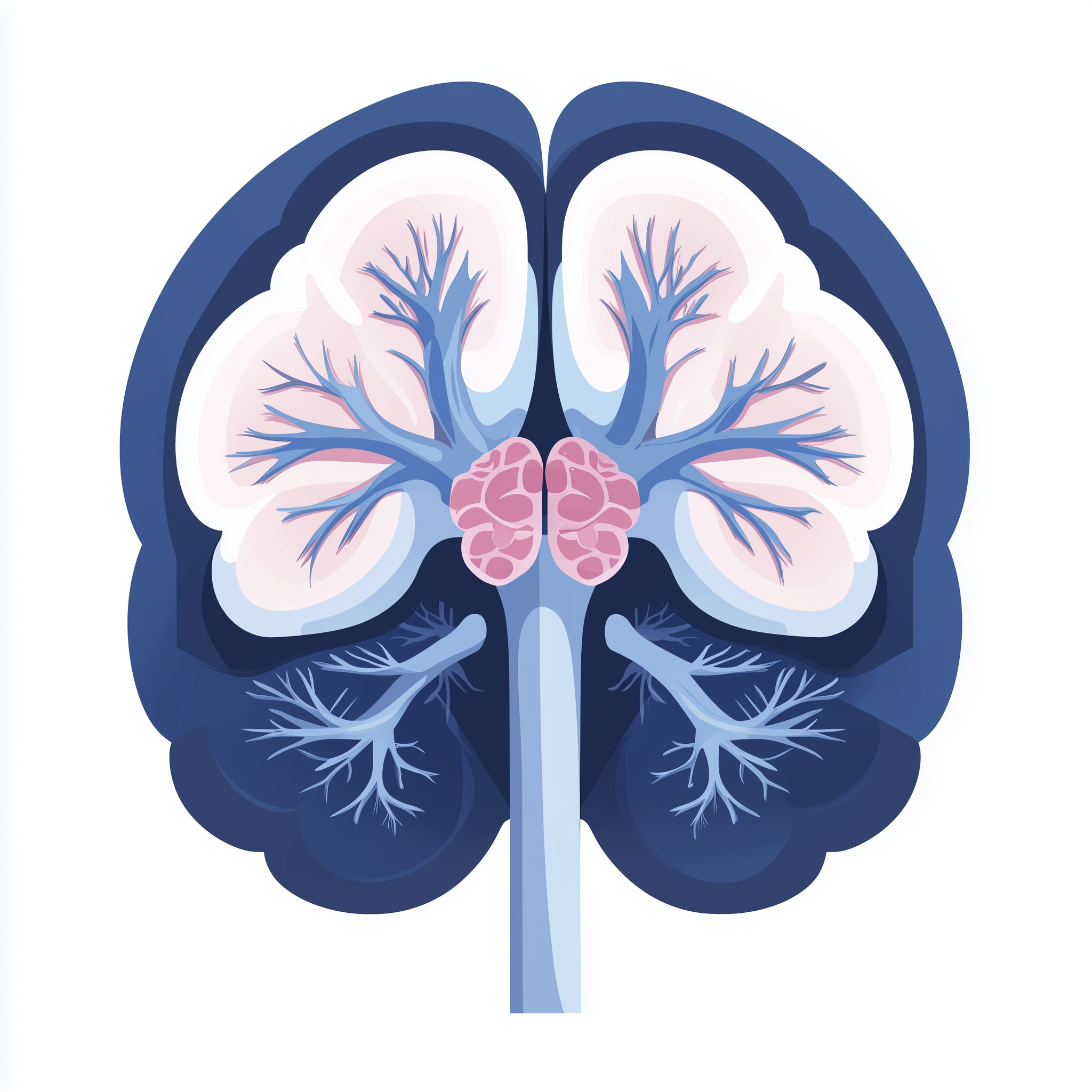
Alexander disease primarily affects the central nervous system, particularly the white matter of the brain. The cerebellum, which coordinates movement and balance, and the brainstem, which controls vital functions such as breathing and heart rate, are often significantly affected in later-onset forms. Characteristic abnormalities can be seen on brain imaging, including changes to specific regions such as the frontal lobes and areas surrounding the fluid-filled spaces (ventricles) within the brain.
The condition also affects the spinal cord in some individuals, particularly those with juvenile or adult-onset disease. Unlike some leukodystrophies, Alexander disease does not typically cause significant problems with organs outside the nervous system, though difficulties with swallowing may lead to nutritional challenges and respiratory complications over time.

Risks & severity
The severity of Alexander disease varies considerably depending on the age of onset. Infantile-onset forms are generally the most severe, with many affected children experiencing significant developmental impairment and shortened life expectancy, often into childhood or adolescence. Juvenile-onset forms, appearing between ages two and twelve years, tend to progress more slowly, and affected individuals may survive into adulthood. Adult-onset Alexander disease is the mildest form, with some individuals maintaining relatively good function for many years after diagnosis, though the condition remains progressive.
Complications can include severe motor disability, nutritional difficulties due to swallowing problems, respiratory infections, and seizures that may be challenging to manage. The progressive nature of the condition means that care needs typically increase over time, requiring multidisciplinary support to maintain quality of life and manage complications as they arise.
Genetic causes
Alexander disease is caused by pathogenic variants in both copies of GFAP in affected individuals. GFAP encodes glial fibrillary acidic protein, which forms a crucial part of the structural scaffold within astrocytes. These star-shaped cells provide essential support to nerve cells throughout the brain and spinal cord. Astrocytes help maintain the chemical environment around nerve cells, regulate blood flow, and contribute to the formation and maintenance of the blood-brain barrier.
When both copies of GFAP carry disease-causing variants, the reduction or absence of functional protein disrupts the normal internal architecture of astrocytes. Loss of adequate GFAP function compromises the structural integrity of these support cells, leading to formation of abnormal protein clumps known as Rosenthal fibres. These aggregates accumulate inside astrocytes and represent a defining microscopic feature of the condition. Because astrocytes are critical for preserving nerve cell health and white matter integrity, their deterioration drives the progressive breakdown of brain white matter and the resulting neurological features seen in affected individuals.
- GFAP glial fibrillary acidic protein
Inheritance pattern
Alexander disease follows an autosomal recessive inheritance pattern. This means that affected individuals have pathogenic variants in both copies of GFAP, one inherited from each parent. Parents of an affected child are typically carriers, meaning they each have one pathogenic variant and one working copy of the gene. Carriers generally do not show symptoms of the condition because one functional copy of GFAP is sufficient for normal astrocyte function.
When both parents are carriers, there is a 25 per cent chance with each pregnancy that the child will inherit both pathogenic variants and be affected by Alexander disease. There is a 50 per cent chance the child will be a carrier like the parents, and a 25 per cent chance the child will inherit two working copies of the gene. Genetic counselling is recommended for families to discuss recurrence risks, carrier testing for relatives, and reproductive options including prenatal diagnosis or preimplantation genetic testing.
When both parents are carriers, each child has a 25% chance of being affected, 50% of being a carrier, and 25% of being unaffected.
Diagnosis & testing
Diagnosis of Alexander disease typically involves a combination of clinical assessment, brain imaging, and genetic testing. Magnetic resonance imaging (MRI) of the brain often reveals characteristic patterns of white matter abnormality and specific changes that can suggest the diagnosis. The combination of clinical features, such as developmental delay with macrocephaly in infantile cases or specific brainstem symptoms in later-onset forms, together with typical MRI findings, may raise suspicion of Alexander disease.
Confirmation requires genetic testing to identify pathogenic variants in both copies of GFAP. Within the NHS, testing for Alexander disease would typically be arranged through clinical genetics services following referral from a paediatric neurologist or adult neurologist. Genetic testing is performed using DNA extracted from a blood sample, and results are usually available within several weeks to months. Once a diagnosis is confirmed in an affected individual, carrier testing may be offered to family members following appropriate genetic counselling.
Management & lifestyle
Management of Alexander disease focuses on addressing symptoms and maintaining quality of life, as there is currently no cure or treatment that can halt disease progression. A multidisciplinary approach involving neurologists, physiotherapists, occupational therapists, speech and language therapists, and dietitians is typically recommended. Antiepileptic medications may be used to manage seizures, though optimal seizure control can be challenging to achieve in some individuals. Physiotherapy and occupational therapy can help maintain mobility and function for as long as possible and prevent complications such as contractures.
Difficulties with swallowing may require assessment by a speech and language therapist, with modifications to food texture and liquid consistency to reduce the risk of aspiration. In some cases, feeding via a gastrostomy tube may be recommended to ensure adequate nutrition and hydration. Respiratory infections should be treated promptly, and some individuals may benefit from physiotherapy techniques to assist with airway clearance. Regular follow-up with a neurologist is important to monitor disease progression and adjust supportive care as needed. Genetic counselling should be offered to families to discuss inheritance, recurrence risks, and reproductive options.
UK care pathway
Within the NHS Genomic Medicine Service, Alexander disease falls under neurogenetics pathways. Referral to clinical genetics services typically occurs following assessment by a neurologist who suspects the diagnosis based on clinical features and brain imaging findings. Genetic testing for GFAP variants would be arranged through regional genetics laboratories as part of the diagnostic pathway for leukodystrophies.
Families affected by Alexander disease have access to genetic counsellors who can explain test results, discuss inheritance patterns, and explore options for future family planning, including prenatal diagnosis or preimplantation genetic testing where appropriate. Ongoing care is coordinated between neurology, genetics, and other specialist services to provide comprehensive support for affected individuals and their families.
Frequently asked questions
How is Alexander disease different from other leukodystrophies?
Alexander disease is distinguished from other leukodystrophies by its cause (pathogenic variants in GFAP rather than genes affecting myelin metabolism), characteristic brain imaging features, and the presence of Rosenthal fibres in brain tissue. The age of onset and specific symptom pattern also help differentiate it from other white matter conditions.
Can Alexander disease be detected before birth?
Yes, when both parental pathogenic GFAP variants are known, prenatal testing can be offered during pregnancy through chorionic villus sampling or amniocentesis. Preimplantation genetic testing may also be available for families considering assisted reproduction. Genetic counselling is recommended to discuss these options.
Is there any treatment that can slow down or stop Alexander disease?
Currently, there is no treatment that can cure Alexander disease or halt its progression. Management focuses on treating symptoms such as seizures, maintaining nutrition and mobility, and preventing complications. Research into potential therapies is ongoing, and families may wish to discuss participation in research studies with their care team.
If both parents are carriers, what is the chance our next child will be affected?
When both parents are carriers of a pathogenic GFAP variant, there is a 25 per cent chance with each pregnancy that the child will inherit both variants and be affected by Alexander disease. There is a 50 per cent chance the child will be a carrier, and a 25 per cent chance the child will inherit two working copies. Genetic counselling can help families understand these risks.
What is the outlook for children diagnosed with Alexander disease?
The outlook varies depending on age of onset. Infantile forms tend to be more severe with significant developmental challenges and shortened life expectancy. Juvenile and adult-onset forms typically progress more slowly, with some individuals maintaining function for many years. A multidisciplinary care team can help optimise quality of life and manage complications.
References
- Messing A. Alexander disease. Handbook of clinical neurology. 2018. PMID: 29478608
- Perlman SJ, Mar S. Leukodystrophies. Advances in experimental medicine and biology. 2012. PMID: 22411242
- Messing A, Brenner M. GFAP at 50. ASN neuro. 2020. PMID: 32811163
- Kuhn J, Cascella M. Alexander Disease. 2023. PMID: 32965913
- Tavasoli A, Armangue T, Ho CY. Alexander Disease. Journal of child neurology. 2017. PMID: 28112050
- Adam MP, Bick S, Mirzaa GM. Alexander Disease. 1993. PMID: 20301351
- Messing A, Brenner M, Feany MB. Alexander disease. The Journal of neuroscience : the official journal of the Society for Neuroscience. 2012. PMID: 22496548
- Hartog L, Dekeyzer S, Bossche SV. Alexander Disease. Journal of the Belgian Society of Radiology. 2021. PMID: 34723085