

On this page
Familial hypomagnesaemia with nephrocalcinosis (CLDN16)
This condition typically presents in infancy or early childhood with low blood magnesium levels despite normal dietary intake. Progressive kidney damage occurs due to calcium deposition in kidney tissue, which can lead to chronic kidney disease if not carefully monitored and managed.
Overview
Familial hypomagnesaemia with nephrocalcinosis (CLDN16) is a rare genetic condition affecting the kidneys' ability to retain magnesium and regulate calcium handling. The disorder arises from pathogenic variants in CLDN16, which disrupt normal kidney tubule function. Children with this condition typically lose excessive amounts of magnesium through their urine, leading to persistently low blood magnesium levels that can affect multiple body systems.
The hallmark feature of nephrocalcinosis - calcium deposits within the kidney tissue itself - distinguishes this condition from simple magnesium deficiency. These deposits form over time and can progressively impair kidney function. Early recognition and intervention are important for preserving kidney health and preventing complications associated with chronic electrolyte imbalance.
Whilst exact prevalence figures are not well established, this condition is recognised as rare, with most cases identified through investigation of unexplained low magnesium levels in childhood or following detection of kidney calcification on imaging studies.
Symptoms & clinical features
The clinical features of familial hypomagnesaemia with nephrocalcinosis typically emerge during infancy or early childhood, though presentation can vary. Affected children may experience muscle cramps, weakness, or abnormal muscle contractions (tetany) due to low magnesium and secondary calcium disturbances. Some infants present with seizures, particularly if magnesium levels drop significantly. Growth may be slower than expected, and recurrent urinary tract infections can occur.
As the condition progresses, kidney-related symptoms become more apparent. Children may produce excessive amounts of urine (polyuria) and experience increased thirst. The calcium deposits within the kidneys often cause no immediate symptoms but contribute to gradual decline in kidney function over months to years. Some affected individuals develop kidney stones, which can cause pain, blood in the urine, or urinary obstruction. Laboratory testing reveals persistently low blood magnesium despite normal or high magnesium in the urine, alongside elevated urinary calcium excretion.
Video: Genetics 101
Affected organs
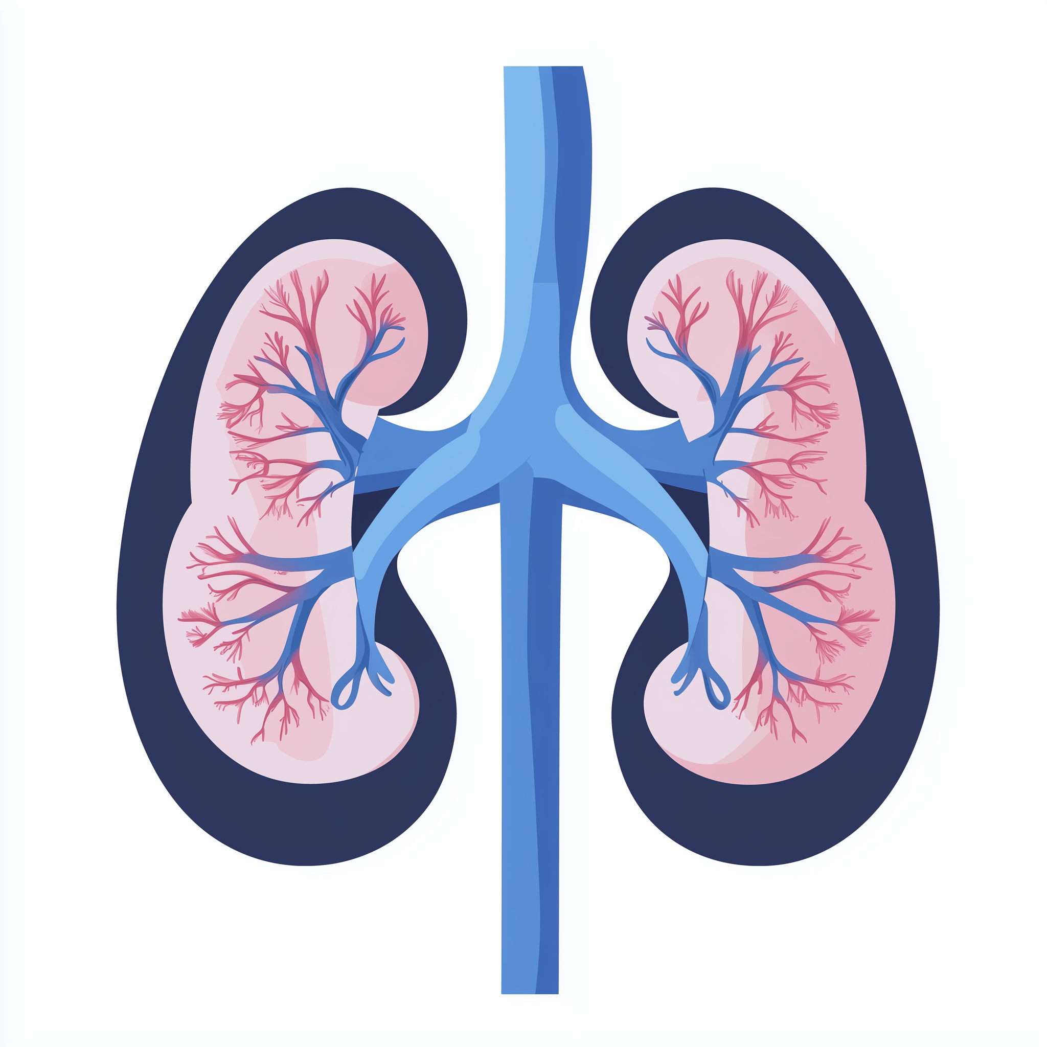
The kidneys are the primary organs affected in this condition. Specifically, the disorder impacts the thick ascending limb of the loop of Henle, a specialised region of the kidney tubule responsible for reabsorbing magnesium and calcium from the forming urine back into the bloodstream. The calcium deposits (nephrocalcinosis) form within the kidney tissue itself, particularly in the medullary region, gradually interfering with normal kidney architecture and function.
Secondary effects can involve other organ systems due to chronic magnesium deficiency. Muscles may be affected, leading to weakness and cramping. The nervous system can be impacted, potentially causing seizures or altered nerve function. Bone health may be compromised over time, as magnesium plays important roles in bone metabolism. The eyes occasionally develop abnormalities, though this is less consistent across affected individuals.

Risks & severity
The severity of familial hypomagnesaemia with nephrocalcinosis varies considerably between individuals, even among those with pathogenic variants in the same gene. Some children experience relatively mild symptoms with stable kidney function when managed appropriately, whilst others develop progressive kidney impairment leading to chronic kidney disease during childhood or adolescence. Research suggests that early diagnosis and consistent management may help slow the rate of kidney function decline, though complete prevention of nephrocalcinosis has proven difficult.
The most significant long-term risk is progression to end-stage kidney disease, which may require dialysis or kidney transplantation. The age at which severe kidney impairment develops is variable, with some individuals maintaining reasonable kidney function into adulthood whilst others experience earlier decline. Additional risks include complications from severe hypocalcaemia (low blood calcium) or hypomagnesaemia, such as cardiac rhythm disturbances or seizures, particularly if electrolyte levels are not adequately monitored and corrected.
Genetic causes
Familial hypomagnesaemia with nephrocalcinosis (CLDN16) results from pathogenic variants in CLDN16, which provides instructions for producing a protein called claudin-16 (also known as paracellin-1). This protein is a critical component of tight junctions between cells lining the thick ascending limb of the kidney tubule. These tight junctions normally act as selective barriers, allowing certain ions to pass between cells whilst preventing others from leaking back into the forming urine.
Claudin-16 works in partnership with other proteins to create a specialised pathway that enables magnesium and calcium to be reabsorbed from the tubular fluid back into the bloodstream. When CLDN16 variants disrupt the normal structure or function of claudin-16, this reabsorption pathway becomes impaired. Consequently, excessive magnesium and calcium are lost in the urine rather than being retained by the body. The high urinary calcium concentration promotes calcium salt precipitation within the kidney tissue, leading to the characteristic nephrocalcinosis. Over time, these deposits and the ongoing electrolyte disturbances contribute to progressive kidney damage.
- CLDN16 claudin 16
Inheritance pattern
Familial hypomagnesaemia with nephrocalcinosis (CLDN16) follows an autosomal recessive inheritance pattern. This means an affected individual has inherited two altered copies of CLDN16 - one from each parent. Parents of an affected child are typically carriers, possessing one pathogenic variant and one functioning copy of the gene. Carriers generally do not show symptoms of the condition, as a single working copy of CLDN16 is sufficient for normal kidney function.
When both parents are carriers, each pregnancy carries a 25 per cent chance of the child being affected, a 50 per cent chance of the child being a carrier like the parents, and a 25 per cent chance of the child inheriting two normal copies of the gene. Siblings of an affected individual have a two in three chance of being carriers if they are unaffected. Genetic counselling can help families understand these inheritance patterns and discuss options for family planning and testing of at-risk relatives.
When both parents are carriers, each child has a 25% chance of being affected, 50% of being a carrier, and 25% of being unaffected.
Diagnosis & testing
Diagnosis of familial hypomagnesaemia with nephrocalcinosis typically begins with recognition of persistent low blood magnesium levels alongside inappropriately high urinary magnesium excretion. Blood tests reveal hypomagnesaemia, often with low blood calcium as well. Urine testing demonstrates elevated magnesium and calcium excretion. Kidney imaging, usually via ultrasound or occasionally CT scanning, identifies nephrocalcinosis as increased echogenicity or calcification within the kidney tissue.
Confirmation requires genetic testing to identify pathogenic variants in CLDN16. Within the NHS Genomic Medicine Service, suspected cases are typically referred to clinical genetics services or specialist paediatric nephrology centres. Testing may be arranged through relevant gene panels for renal tubular disorders. Families concerned about this condition should discuss referral with their GP or paediatric specialist, who can coordinate appropriate investigations and onward referral to genetic services where genetic counselling and testing can be accessed.
Management & lifestyle
Management of familial hypomagnesaemia with nephrocalcinosis focuses on maintaining electrolyte balance and preserving kidney function for as long as possible. Affected individuals typically require lifelong magnesium supplementation, often in substantial doses, though complete normalisation of blood magnesium may be difficult to achieve. Additional supplementation with potassium or other electrolytes may be necessary depending on individual biochemical profiles. Regular monitoring of kidney function, electrolyte levels, and kidney imaging is essential to track disease progression and adjust treatment accordingly.
Some individuals benefit from thiazide diuretics, which can reduce urinary calcium excretion and potentially slow the progression of nephrocalcinosis, though evidence for long-term benefit remains limited. Adequate hydration is generally encouraged. If kidney function declines significantly, management strategies for chronic kidney disease become necessary, potentially including dietary modifications, treatment of anaemia, and management of bone mineral metabolism. Advanced kidney disease may ultimately require dialysis or kidney transplantation. Care is typically coordinated through specialist paediatric or adult nephrology services within the NHS, with input from clinical genetics and specialist dietitians as needed.
UK care pathway
Within the NHS, individuals with suspected familial hypomagnesaemia with nephrocalcinosis are typically identified through paediatric nephrology or general paediatric services when investigations reveal unexplained hypomagnesaemia or nephrocalcinosis. Referral to specialist paediatric nephrology centres is standard for ongoing management and monitoring. Genetic testing is accessed through the NHS Genomic Medicine Service, with appropriate gene panels available for inherited renal tubular disorders. The relevant PanelApp panels include genes associated with renal tubular dysfunction and electrolyte disorders.
Families benefit from access to clinical genetics services, where genetic counsellors can explain inheritance patterns, discuss implications for family members, and coordinate predictive or carrier testing for relatives where appropriate. Transition from paediatric to adult nephrology services should be planned carefully to ensure continuity of specialist care. Multidisciplinary input often involves nephrologists, clinical geneticists, specialist nurses, and dietitians working together to optimise long-term outcomes.
Frequently asked questions
Can my child's magnesium levels be completely normalised with supplements?
Complete normalisation of blood magnesium levels is often difficult to achieve, even with high-dose supplementation. Treatment aims to raise magnesium to safer levels and reduce symptoms, but some degree of hypomagnesaemia often persists because the underlying kidney defect continues to cause urinary magnesium loss.
Will the calcium deposits in the kidneys go away with treatment?
Unfortunately, nephrocalcinosis tends to be progressive despite treatment. Current therapies focus on slowing further calcium deposition and preserving remaining kidney function rather than reversing existing calcification. Some interventions may reduce the rate of new deposit formation.
What are the chances my other children are affected or carriers?
If both parents are carriers, each child has a 25 per cent chance of being affected, a 50 per cent chance of being a carrier, and a 25 per cent chance of inheriting two normal gene copies. Genetic counselling can discuss testing options for siblings and other family members.
How often does this condition lead to kidney failure?
The progression to severe kidney impairment varies considerably between individuals. Some maintain reasonable kidney function into adulthood with careful management, whilst others experience earlier decline requiring dialysis or transplantation during childhood or young adulthood. Regular monitoring helps identify changes in kidney function early.