

On this page
Arrhythmogenic right ventricular cardiomyopathy
ARVC is a genetic condition primarily affecting the right ventricle of the heart, leading to problems with heart rhythm and function. It can cause palpitations, fainting, and in some cases, sudden cardiac arrest, often impacting younger individuals and athletes.
Overview
Arrhythmogenic right ventricular cardiomyopathy (ARVC) is a hereditary heart muscle disease that mainly affects the right ventricle, one of the heart's lower chambers. In ARVC, healthy heart muscle tissue gradually degenerates and is replaced by fatty and fibrous tissue [PMID:33675034]. This change in the heart muscle can disrupt the heart's electrical signals, leading to abnormal heart rhythms (arrhythmias). These arrhythmias can range from mild palpitations to life-threatening events such as sudden cardiac arrest.
ARVC is considered a progressive condition, meaning its effects can worsen over time. While it primarily affects the right ventricle, some individuals may also experience involvement of the left ventricle. The condition is often diagnosed in adolescents and young adults, but symptoms can emerge at any age. Its prevalence is estimated to be between 1 in 2,000 and 1 in 5,000 people in the general population, although these figures may vary [PMID:28797457].
Understanding ARVC is crucial for timely diagnosis and management, as early intervention can help prevent serious complications. The condition is typically inherited, meaning it can run in families, highlighting the importance of family screening once a diagnosis is made.
Symptoms & clinical features
The symptoms of ARVC can vary widely among individuals, even within the same family. Some people may experience no symptoms for many years, while others may have severe issues early in life. The most common symptoms are related to the heart's electrical instability and include palpitations, which are sensations of a racing or fluttering heart [PMID:28797457].
Other potential symptoms include dizziness or lightheadedness, particularly during physical activity. Fainting (syncope) can also occur, often due to significant arrhythmias. In some cases, the first sign of ARVC may be sudden cardiac arrest, especially during strenuous exercise. Less commonly, individuals might experience chest pain or shortness of breath, particularly as the disease progresses and affects the heart's ability to pump blood effectively.
Symptoms often become more apparent or worsen during physical exertion, as exercise can trigger arrhythmias in affected individuals. It is important for individuals experiencing these symptoms to seek medical attention, especially if there is a family history of heart conditions or sudden unexplained deaths.
Video: Genetics 101
Affected organs
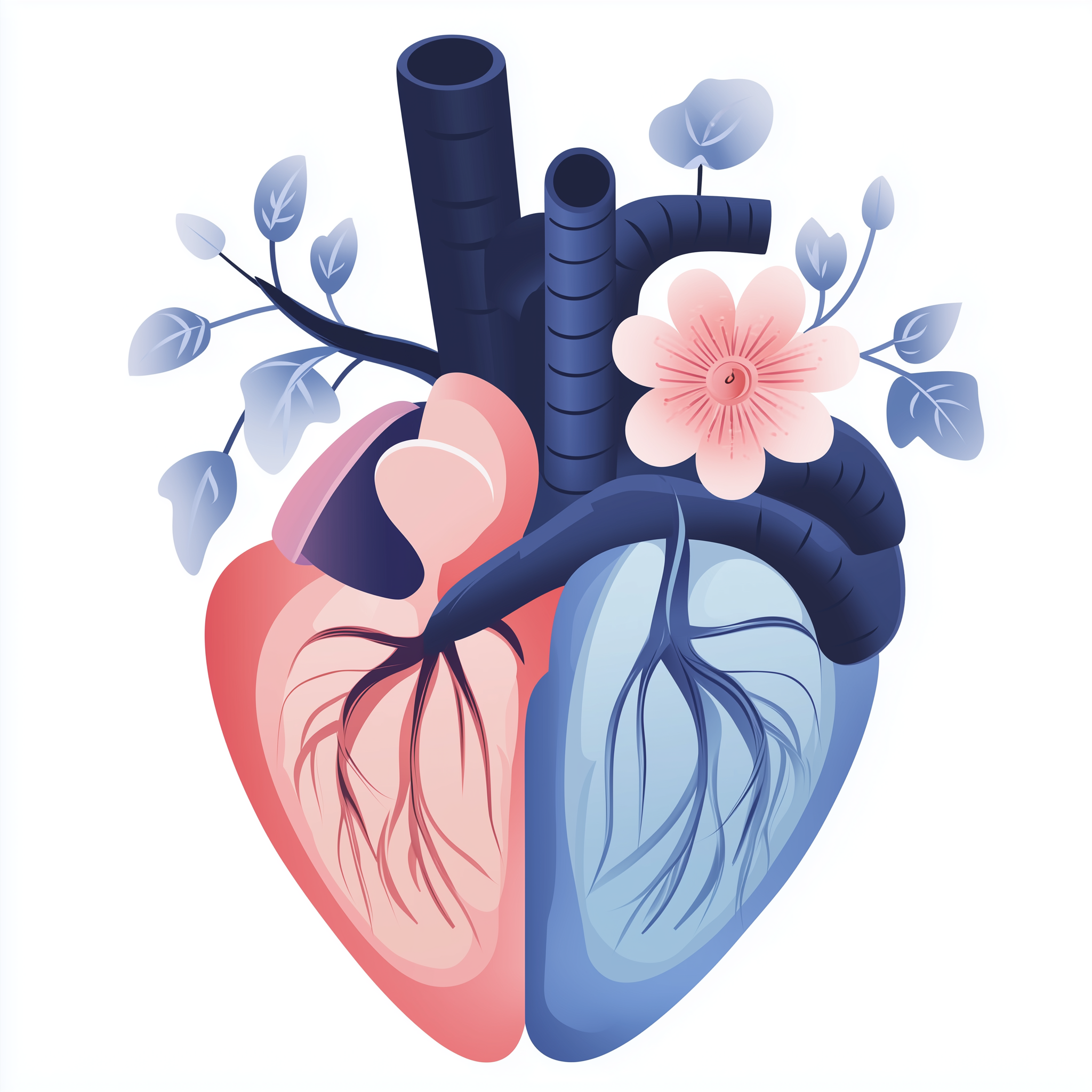
The primary organ affected by ARVC is the heart, specifically the right ventricle. This chamber is responsible for pumping deoxygenated blood to the lungs. In ARVC, the muscle tissue of the right ventricle is progressively replaced by fatty and fibrous tissue. This structural change weakens the heart muscle and makes it prone to electrical instability, leading to arrhythmias.
While the right ventricle is predominantly affected, some individuals with ARVC may also show signs of left ventricular involvement, either in isolation or in combination with right ventricular disease. This can further impair the heart's overall pumping function. The electrical system of the heart is also significantly affected, as the abnormal tissue disrupts the normal conduction of electrical impulses, causing irregular heartbeats. There are no other major organ systems typically affected in ARVC.

Risks & severity
ARVC has a variable clinical course and severity, meaning that the impact of the condition can differ significantly from person to person. The condition is progressive, and the extent of heart muscle damage can increase over time. One of the most serious risks associated with ARVC is sudden cardiac death, particularly in young adults and athletes, often during physical exertion. ARVC is recognised as a leading cause of sudden cardiac death in individuals under 35 years old [PMID:33675034].
The age of onset for symptoms can vary widely, from childhood to older adulthood, but typically symptoms manifest in adolescence or young adulthood. Lifetime risks associated with ARVC include the development of heart failure, where the heart becomes unable to pump enough blood to meet the body's needs, and the need for interventions such as implantable cardioverter-defibrillators (ICDs) to manage life-threatening arrhythmias. Regular monitoring and appropriate management are crucial to mitigate these risks and improve long-term outcomes for individuals with ARVC.
Genetic causes
ARVC is primarily a genetic condition, meaning it is caused by changes (variants) in specific genes. Most of the genes associated with ARVC encode proteins that are vital components of desmosomes. Desmosomes are specialised cell junctions that act like 'spot welds', holding cardiac muscle cells together and enabling them to withstand mechanical stress [PMID:28797457].
The main genes implicated in ARVC include *DSG2*, *DSP*, and *PKP2*. Pathogenic variants in *DSG2* can lead to a dysfunctional desmoglein 2 protein, compromising the structural integrity of desmosomes. Similarly, variants in *DSP* affect desmoplakin, another critical desmosomal protein. The *PKP2* gene encodes plakophilin 2, also a key desmosomal component; changes in this gene are a common cause of ARVC [PMID:28797457]. When these desmosomal proteins are faulty, the heart muscle cells cannot properly connect, leading to cell death and replacement by fibrous and fatty tissue. This process underlies the structural and electrical problems seen in ARVC.
- DSG2 desmoglein 2DSG2 encodes desmoglein 2, a structural protein essential for maintaining cell-to-cell adhesion in heart muscle and other tissues.
- DSP desmoplakinThe DSP gene provides instructions for making desmoplakin, a key protein in desmosomes that helps cells adhere to each other, particularly in the heart and skin.
- PKP2 plakophilin 2The PKP2 gene provides instructions for making plakophilin 2, a crucial protein for the structural integrity of heart muscle cells, particularly within desmosomes.
Inheritance pattern
ARVC is typically inherited in an autosomal dominant pattern. This means that a person only needs to inherit one copy of the altered gene from one parent to develop the condition. If a parent has ARVC, there is a 50% chance with each pregnancy that their child will inherit the pathogenic gene variant and potentially develop the condition.
Since ARVC is autosomal dominant, it affects males and females equally. However, the expression of the condition can vary, meaning that even within the same family, individuals who inherit the gene variant may experience different symptoms or have varying degrees of severity. This is known as variable expressivity. Genetic counselling is highly recommended for families where ARVC has been diagnosed to understand the inheritance pattern and discuss implications for other family members.
Each child has a 50% chance of inheriting the pathogenic variant, regardless of sex.
Diagnosis & testing
The diagnosis of ARVC involves a combination of clinical evaluation, imaging tests, and genetic testing. Clinical suspicion often arises from symptoms such as palpitations, fainting, or a family history of ARVC or sudden cardiac death. Initial investigations may include an electrocardiogram (ECG) to check the heart's electrical activity and an echocardiogram to visualise the heart's structure and function.
More advanced imaging techniques, such as cardiac magnetic resonance imaging (MRI), are often used to detect the characteristic fatty and fibrous replacement of heart muscle in the right ventricle [PMID:33675034]. Diagnosis is typically guided by established clinical criteria, such as the revised Task Force Criteria. Genetic testing is a crucial component of diagnosis, particularly in confirming the underlying cause and for family screening. In the UK, genetic testing for ARVC is available through the NHS Genomic Medicine Service (GMS) pathways, often initiated following referral to a clinical genetics service or specialist cardiology clinic, with relevant R-codes for testing. This helps to identify specific pathogenic variants in genes like *DSG2*, *DSP*, and *PKP2*.
Management & lifestyle
Managing ARVC focuses on controlling symptoms, preventing life-threatening arrhythmias, and slowing the progression of heart disease. Treatment strategies are individualised based on the severity of the condition and the patient's specific symptoms. Lifestyle modifications, such as avoiding strenuous exercise, are often recommended, as physical activity can trigger arrhythmias in individuals with ARVC.
Medications such as beta-blockers may be prescribed to help manage heart rhythm and reduce the risk of arrhythmias. For individuals at high risk of sudden cardiac death, an implantable cardioverter-defibrillator (ICD) may be recommended. An ICD is a small device surgically placed in the chest that can detect and deliver an electrical shock to correct dangerous heart rhythms [PMID:28797457]. Regular follow-up appointments with a cardiologist are essential to monitor heart function and adjust management plans as needed. The NHS provides comprehensive care for individuals with ARVC, typically through specialist cardiology centres with access to multidisciplinary teams.
UK care pathway
In the UK, individuals suspected of having ARVC are typically referred to specialist cardiology services or clinical genetics departments. These services are part of the NHS Genomic Medicine Service (GMS), which provides advanced diagnostic testing and expert care. Genetic testing for ARVC is available through the GMS, with specific R-codes used to guide testing based on clinical need and family history.
Following a diagnosis of ARVC, patients often receive ongoing care from a multidisciplinary team, including cardiologists, electrophysiologists, and genetic counsellors. Genetic counsellors play a crucial role in explaining the inheritance pattern, discussing the implications for family members, and arranging cascade screening where appropriate. This integrated approach ensures that patients and their families receive comprehensive support and management for this complex condition.
Frequently asked questions
Can ARVC be prevented?
ARVC is a genetic condition, so it cannot be prevented. However, early diagnosis and appropriate management can help prevent serious complications and improve quality of life. For family members of someone with ARVC, genetic counselling and screening can identify those at risk.
Is ARVC always severe?
No, the severity of ARVC can vary greatly among individuals, even within the same family. Some people may have mild symptoms or none at all for many years, while others may experience more significant heart rhythm problems or heart failure. Regular monitoring helps to manage the condition.
Can I exercise if I have ARVC?
Generally, individuals with ARVC are advised to avoid strenuous exercise and competitive sports, as physical activity can trigger dangerous heart rhythms. Your cardiologist will provide specific guidance based on your individual condition and risk profile.
How often do I need to be monitored if I have ARVC?
Regular monitoring by a cardiologist is crucial for individuals with ARVC. The frequency of appointments and tests, such as ECGs and echocardiograms, will depend on your symptoms, the severity of your condition, and your individual risk factors. Your care team will create a personalised follow-up plan.
What is the role of genetic testing in ARVC?
Genetic testing can confirm a diagnosis of ARVC by identifying specific genetic changes associated with the condition. It is also very important for family members, as it can help identify those who have inherited the genetic change and may be at risk, allowing for early monitoring and intervention.
References
- Corrado D, Link MS, Calkins H. Arrhythmogenic Right Ventricular Cardiomyopathy. The New England journal of medicine. 2017. PMID: 28052233
- Tsatsopoulou A. Arrhythmogenic right ventricular cardiomyopathy and pregnancy. International journal of cardiology. 2018. PMID: 29544930
- Brugada J, Hindricks G. Primary electrical disorders and arrhythmogenic right ventricular cardiomyopathy: new research insights with clinical implications. Europace : European pacing, arrhythmias, and cardiac electrophysiology : journal of the working groups on cardiac pacing, arrhythmias, and cardiac cellular electrophysiology of the European Society of Cardiology. 2018. PMID: 29878208
- Corrado D, Zorzi A. Arrhythmogenic right ventricular cardiomyopathy and sports activity. European heart journal. 2015. PMID: 25971286
- Cheng WH, Chung FP, Lin YJ. Arrhythmogenic right ventricular cardiomyopathy: diverse substrate characteristics and ablation outcome. Reviews in cardiovascular medicine. 2021. PMID: 34957771
- Kaddoura R, Al-Tamimi H. Physical Exercise and Arrhythmogenic Right Ventricular Cardiomyopathy/Dysplasia: An Overview. Heart views : the official journal of the Gulf Heart Association. 2022. PMID: 36605925
- Alblaihed L, Kositz C, Brady WJ. Diagnosis and management of arrhythmogenic right ventricular cardiomyopathy. The American journal of emergency medicine. 2023. PMID: 36638611
- Adam MP, Bick S, Mirzaa GM. Arrhythmogenic Right Ventricular Cardiomyopathy Overview. 1993. PMID: 20301310